Video-Assisted Thoracic Surgery: Minimally Invasive Surgery for Myasthenia Gravis (MG)

Muscle fatigue is a typical symptom that decreases muscles’ ability to perform over time. It can be temporarily associated with a state of exhaustion, often following strenuous activities or exercise. However, if muscle fatigue persists or progresses from localized to generalized weakness, it might indicate the abnormalities of nerves and muscles (neuromuscular system) which is known as “Myasthenia Gravis” or shortly called “MG”.
Get to know MG
Myasthenia Gravis (MG) is defined as a long-term neuromuscular disease that leads to varying degrees of muscle weakness. It is characterized by rapid fatigue or weakness of any muscles under voluntary control which is caused by a breakdown in the regular communication between nerves and muscles. In normal pathway, nerves communicate with muscles by releasing neurotransmitters (chemical substances) called “acetylcholine”. In myasthenia gravis, immune system (particularly the thymus gland) produces abnormal proteins, known as “antibodies” that block or destroy acetylcholine. As a result, muscles receive fewer nerve signals, eventually cause weakness with different degrees of severity, depending on affected sites.
The estimated prevalence of myasthenia gravis is approximately 3 cases per 100,000 population. This disease affects all age groups, ranging from children to the elderly. In fact, myasthenia is found predominantly in adolescent women and elderly men.
Fluctuating muscle weakness and abnormal fatigability vary among individual patients. Although myasthenia gravis can affect any of the voluntary muscles, certain muscle groups are more commonly affected including eye muscles, face and throat muscles as well as neck and limb muscles. If myasthenia gravis affects eyes muscles, related symptoms might include drooping of one or both eyelids (ptosis) or/and double vision (diplopia). If muscles of face and throat are impacted, manifested symptoms are impaired speaking with nasal sound, difficulty swallowing and choking. Myasthenia gravis can also further cause weakness in the neck, arms and legs, leading to walking difficulties. In addition, medical emergency condition “myasthenic crisis” leads to acute respiratory failure which is defined as a sudden malfunction in the ability of respiratory system to maintain adequate gas exchange. In such a case, intubation and mechanical ventilation are essentially required.
Generally, muscle weakness caused by myasthenia gravis can come and go. However, symptoms get normally worse when affected muscle is used. On the contrary, related symptoms usually improve with rest.
Nevertheless, these symptoms might be relatively associated with other diseases, for instance, myositis (inflammation of the muscles), nerves diseases, Motor Neurone Disease (MND) such as amyotrophic lateral sclerosis and other neurodegenerative disorders. Confirmative diagnostic tests of myasthenia gravis include:
- Neurological examination and tests to determine neurological abnormalities including reflexes, muscle strength and tone, senses of touch and sight, coordination and balance.
- Repetitive nerve stimulation. Electrodes are attached to the skin over the muscles to be tested. Small pulses of electricity is sent through the electrodes to measure the nerve’s ability to send a signal to muscles.
- Blood analysis to determine the presence of abnormal antibodies that disrupt nerve signals.
- Ice pack test. For droopy eyelid, a bag filled with ice will placed on the eyelid for few minutes. After removal of the ice pack, the signs of improvement will be further evaluated.
- Edrophonium test: Injection of the edrophonium (to block enzyme that breaks down acetylcholine) that results in a sudden, temporary improvement in muscle strength might indicate myasthenia gravis.
- Imaging tests such as abdominal CT scan or MRI might be additionally required to further investigate the abnormalities in the thymus glands. Investigations of thyroid and other auto-immune diseases might be also needed.
Treatment
If diagnosed promptly, myasthenia gravis can be successfully treated. The effectiveness of treating depends primarily on the severity of the disease, the duration of the disease, disease progression, the patient’s age and overall health. Treatment of myasthenia gravis consists of 2 main approaches; medications and surgery.
- Medications
Myasthenia gravis can be well-controlled with the following medications:
- Cholinesterase inhibitors. These medicines are commonly used drugs to treat myasthenia gravis but they are most useful in mild forms of the condition such as drooping of one or both eyelids. They enhance communication between nerves and muscles by preventing the breakdown of acetylcholine, thus increasing the muscle’s ability to contract. Although they cannot cure the disease, they substantially help improving muscle contraction and muscle strength. The most commonly prescribed form of this drug is pyridostigmine, under a trade name “Mestinon®”.
- Immunosuppressants: For more severely ill patients, corticosteroids and immunosuppressants such as cyclosporine and azathioprine help suppressing the activity of the immune system. Although they are often very effective with high-dose corticosteroid regimen, these drugs typically have serious associated side effects e.g. increased risk of infection and liver or kidney damage. Despite their efficacy, steroid-induced side effects generally require tapering of the drug as soon as the symptoms of myasthenia gravis are under control. Moreover, tapering must be done carefully to avoid uncontrolled myasthenia gravis. These drugs cannot be stopped abruptly since it might aggressively aggravate symptoms including difficulty breathing, difficulty swallowing, choking and respiratory failure which intubation and mechanical ventilation are usually required. More importantly, potential advantages versus risks of taking these drugs should be clearly discussed with specialized doctors.
- Intravenous immunoglobulin (IVIg): This therapy modulates the immune system and reduces the effects of causative autoantibodies. It is usually used in the short term to treat a sudden worsening of symptoms, especially with breathing involvement or before other therapies for long-term disease management.
- Surgery
The thymus gland is one of the endocrine glands and also a lymphoid organ of the immune system. This small flattened organ in the shape of a butterfly is situated in the upper chest beneath the sternal bone (breastbone). The thymus gland serves a vital role in the development of T-lymphocytes or T cells, an extremely important type of white blood cell. It also produces thymosin, hormone that stimulates the development of disease-fighting T cells. Typically, the thymus gland begins to grow at birth and continues until puberty. After puberty, the thymus starts to slowly shrink and become replaced by fat.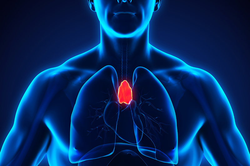
Role of the thymus gland in myasthenia gravis
Myasthenia gravis is a chronic autoimmune neuromuscular disease that causes weakness in the skeletal muscles. The thymus gland plays a crucial role in the development of immune system, particularly the production of T cells. Clinical studies indicate that removing the gland improves myasthenia gravis symptoms since this gland triggers the production of the antibodies that block neurotransmitter “acetylcholine”, resulting in impaired nerve signals and leading to different degrees of muscle weakness. In 2016, the clinical trials were conducted in order to compare the treatment effectiveness between medication treatment (as a monotherapy) and surgical removal of the thymus gland (thymectomy). The results revealed that surgical treatment could improve patients’ weakness with significant reduction in medication needs such as immunosuppressants therefore leading to less serious side effects
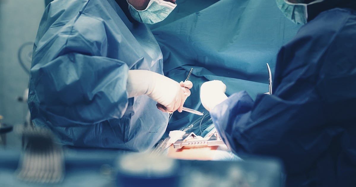
Surgical removal of the thymus gland
To surgically remove the thymus gland, a thymectomy can be performed as an open surgery or as a minimally invasive surgery:
- Open surgery:
In this conventional procedure, a considerably large incision is made in the skin over the breastbone (sternum) and the breastbone is divided or cracked to expose the thymus (called median sternotomy). Then surgeon removes the thymus through this open cut to achieve adequate resection margins and clearance of the mediastinal fat. This approach was commonly used in the past for surgical resection of the thymus gland. - Minimally Invasive Surgery: Video-Assisted Thoracic Surgery or VATS
In recent years, VATS thymectomy has been gaining acceptance due to its superior advantages. It is a minimally invasive surgery to remove the thymus gland through smaller incisions. Surgeons make few small incisions in the side of the chest. Small surgical instruments are inserted to visualize and remove the thymus gland. Instead of having a large cut required in the open surgery, VATS does not need breastbone to be divided in order to remove the thymus. As a result, small incisions can significantly lead to less pain, enhanced cosmetic benefit and fewer post-operative complications as well as faster recovery time and shorter hospital stay that generally takes only 1-3 days, whereas open surgery takes 5-7 days. Compared to conventional approach, patients can return to their daily life and activities even quicker.
Multiportal VATS
The conventional VATS involves 3-4 small incisions (called conventional multiportal VATS). This approach typically consists of three ports which are small incisions of 3–4 cm. long. The first incision is used to insert video camera called a thoracoscope, a thin, flexible tube with a light and a small video camera on the end. The second and third incisions accommodate other surgical instruments such as retractors, clamps and cutting equipment. The position of video camera is normally placed between intercostal spaces, the 7th – 8th ribs and the location of other ports for surgical instrument is between the spaces of the 4th – 6th ribs, both posterior and anterior sites.
Although VATS involves smaller skin incisions and it avoids the use of rib retractors, trocar insertions are essentially required at several intercostal spaces. It is subsequently predisposed to causing intercostal nerve injury at several sites around these small incisions. Trocar and other surgical instrument insertions might potentially damage surrounding nerves with different degrees of severity, resulting in post-operative pain at the surgical sites.
Uniportal VATS
Uniportal VATS has been generating an increasing interest due to promising superior results compared to conventional multiportal VATS. Only one 3–4 cm incision is necessary (between 4th or 5th intercostal space) with no muscle and nerve disruptions, no rib spreading and no necessity of trocars. A long thoracoscope is used, hold in the upper part of the incision and all the other instruments can be inserted in the lower part of the same incision. Uniportal VATS gives the possibility of introducing several instruments through the same small incision and handling them comfortably and safely. However, this advanced procedure needs highly skilled and experienced surgeons who are specialized in multiportal VATS (triportal and biportal VATS). Its major benefit, compared to multiportal VATS, is the reduction of pain with greater satisfaction from a single incision while the clinical outcomes remain equivalent.
Lifestyle and home remedies
Since myasthenia gravis is a chronic condition, doctor’s recommendations must be followed strictly to achieve the best possible treatment outcomes and reduce chances of serious complications. Most cases of patients diagnosed with myasthenia gravis are prescribed corticosteroid drugs to suppress immune activities. Common side effects of long-term corticosteroid drugs include:
- An increased appetite, leading to weight gain. It is highly advised to restrict high-calorie diet intake in order to prevent the development of other health problems such as obesity and diabetes.
- An increased susceptibility to several different types of infections. Direct exposure to infectious persons must be avoided. Annual flu vaccination is advised collectively with other necessary vaccines.
- Other undesired side effects are high blood pressure and steroid induced glaucoma and cataract. Close monitoring is virtually required.
- In case that steroid is prescribed for long-term treatments, calcium and vitamin D supplements should be concurrently given in order to prevent steroid related osteoporosis.
- Home remedies include regular exercise, weight control and adjusting eating routine e.g. consumption of healthy diets with low calories such as fruits and vegetables.
References:
1. Gil I. Wolfe et al, Randomized Trial of Thymectomy in Myasthenia Gravis. N Engl J Med 2016; 375:511-522
2. Shen Y et al. Single versus multiple-port thoracoscopic lobectomy for lung cancer: a propensity-matched study. Eur J Cardiothorac Surg. 2016 Jan;49 Suppl 1:i48-53
3. Xu GW et al .A prospective comparative study examing the impact of uniportal and three portal video-assisted thoracic surgery on short-term quality of life in lung cancer. Zhonghua Wai Ke Za Zhi. 2018 Jun 1;56(6):452-457
4. Mu JW et al. A Matched Comparison Study of Uniportal Versus Triportal Thoracoscopic Lobectomy and Sublobectomy for Early-stage Nonsmall Cell Lung Cancer. Chin Med J (Engl). 2015 Oct 20;128(20):2731-5.
5. Young R et al. Is uniport thoracoscopic surgery less painful than multiple port approaches? Interact Cardiovasc Thorac Surg. 2015 Mar;20(3):409-14