“Pneumothorax” a medical urgency that has to be treated

A pneumothorax (collapsed lung) occurs when air leaks into the space between the lung and chest wall. This air pushes on the outside of the lung and makes the lung collapse. Pneumothorax can be a complete lung collapse or a collapse of only a portion of the lung. Pneumothorax can attack anyone at anytime. However, high risk group to develop pneumothorax include the elderly who are heavy smokers, working people who expose to air pollution and adolescent group with precocious puberty. Pneumothorax can be caused by a blunt or penetrating chest injury, certain medical procedures or damage from underlying lung disease. It might be silent without manifestation of any sign. Typical symptoms are sudden chest pain and shortness of breath accompanied with dry cough. If left untreated, pneumothorax can be a life-threatening condition.
Get to know pneumothorax
A pneumothorax (collapsed lung) occurs when air leaks into the space between the lung and chest wall. This air pushes on the outside of the lung and makes the lung collapse. Pneumothorax causes leaking air to accumulate between the chest wall and the lung and increases pressure in the chest, reducing the amount of blood returned to the heart. Symptoms include chest pain, shortness of breath, rapid breathing and a racing heart, followed by shock. Pneumothorax can be a complete lung collapse or a collapse of only a portion of the lung. Since collapsed lung can put pressure on the heart, it is a fatal situation that has to be treated immediately upon diagnosis.
Warning signs of pneumothorax
A pneumothorax can be caused by lung diseases that damage lung tissue such as chronic obstructive pulmonary disease (COPD), cystic fibrosis and pneumonia. Chest injury including any blunt or penetrating injury to the chest can cause lung collapse. Some injuries may happen during physical assaults, car crashes or being stabbed in the chest, while others may inadvertently occur during medical procedures that involve the insertion of a needle into the chest. A severe type of pneumothorax can also occur in people who need mechanical assistance to breathe. The ventilator can create an imbalance of air pressure within the chest. The lung may collapse completely. Signs and symptoms of pneumothorax in different groups vary depending on age, individual health conditions and risk factors.
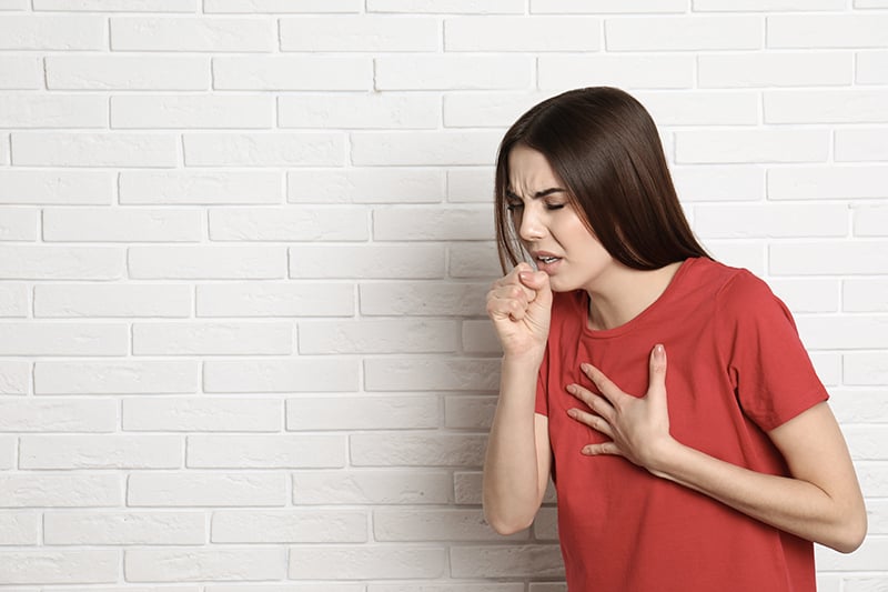
- In young patients: Common signs and symptoms are sudden chest pain accompanied with dry cough without the presence of having previous common cold, runny nose or sore-throat.
- In elderly patients or heavy smokers: Typical symptom is shortness of breath, especially in patients diagnosed with Chronic Obstructive Pulmonary Disease or COPD such as emphysema in which the air sacs in the lungs (alveoli) are damaged.
A sudden chest pain caused by pneumothorax differs from chest pain induced by cardiac diseases. The former produces a sudden sharp chest pain, in particular side of the lung followed by pains during breathing in while the latter causes the feeling like pressure or squeezing in the chest. Complications caused by pneumothorax is fatal, therefore immediate treatment is required.

Types of pneumothorax
There are 2 common types of pneumothorax:
- Primary Spontaneous Pneumothorax (PSP)
Primary spontaneous pneumothorax (PSP) occurs in patients without underlying lung disease and in the absence of an inciting event. Air enters into the intrapleural space between the chest wall without preceding trauma and underlying history of clinical lung disease. Patients are typically aged 18-30 years, tall and thin. Although the cause of primary spontaneous pneumothorax remains unknown, many patients whose condition is labeled as primary spontaneous pneumothorax have certain risk factors including genetic abnormalities and adolescent group with precocious puberty, affecting lung function. In addition, high risk group also includes women with endometriosis, a condition where the endometrium which is the inner uterine lining grows outside of the uterus in areas such as the ovary, pelvic area and intestine. Catamenial pneumothorax is a condition of air leaking into the pleural space occurring in conjunction with menstrual periods or during ovulation, believed to be caused primarily by endometriosis of the pleura (the membrane surrounding the lung or diaphragm). Treatment of catamenial pneumothorax mainly involves surgery and hormonal therapy. - Secondary Spontaneous Pneumothorax (SSP)
Secondary spontaneous pneumothorax (SSP) occurs in patients with a wide variety of lung diseases such as chronic obstructive pulmonary disease (COPD), heavy smokers and the elderly aged over 60. These individuals have underlying pulmonary pathology that alters normal lung structure. Air enters the pleural space via distended, damaged or compromised alveoli, tiny air sacs in the lung. Compared to Primary Spontaneous Pneumothorax, the presentation of these patients may include more serious clinical symptoms and sequelae due to comorbid conditions, particularly in patients with damaged lung tissue or impaired lung function.
In Thailand, Secondary Spontaneous Pneumothorax is more common. Apart from heavy smoking, other possible risk factor might involve extreme air pollution which potentially increases risk of developing emphysema and other lung conditions. Especially in Bangkok and surrounding areas, a cloud of ultra-fine dust particles known as PM2.5 has recently returned. According to the Pollution Control Department (PCD), the concentration of PM2.5 pollutants should not exceed the PCD’s safe threshold of 50 µg/m³. If the number is greater than this threshold level, these fine particulate matters can substantially cause a wide range of health problems including respiratory disease and heart disease. For instance, a middle-aged patient who had run a marathon for 5 consecutive years developed pneumothorax. His imaging results reveal the blackened lungs even though he has never smoked. Normally it takes up to 10 years to manifest relevant signs and symptoms. However, in this case, it showed up extremely quick. In other case, young patient suddenly developed dry cough with severe chest pain during participating in a concert. Diagnostic result indicates pneumothorax. Undiagnosed chronic emphysema was triggered by the loud noise in the concert. As a result, the alveoli became ruptured in the presence of sudden sharp pain in the chest.

Diagnosis of pneumothorax
Pneumothorax is generally diagnosed by using a chest X-ray. In some cases, a computerized tomography (CT) scan may be needed to provide more-detailed images. Ultrasound imaging also may be used to identify a pneumothorax.
Treatment of pneumothorax
Most cases who have pneumothorax attacks seek medical attention in emergency department due to sudden chest pain and shortness of breath. Imaging results obtained from chest X-ray might indicate the air leakage in the space between the lung and chest wall while leakage of the lungs could not be revealed. The goal in treating a pneumothorax is to relieve the pressure on the lung, allowing it to re-expand. Depending on the cause of the pneumothorax, a second goal may be to prevent recurrences. The selected treatment for achieving these goals depend on the severity of the lung collapse and overall health conditions. The previous treatment guideline in the past mainly involved chest tube insertion to reduce pressure on the lung. The leakage could be healed and closed automatically within 3-7 days. Surgery is only indicated in case of recurrence. However, the recurrent rate is considerably high, up to 30% after the first attack.
Due to the advancements in cardiothoracic surgery, Video-Assisted Thoracic Surgery or VATS, a minimally invasive surgery has developed. This minimally invasive technique has been increasingly used due to its superiority including smaller incisions (similar size compared to incision needed for chest tube insertion), less pain and lower postoperative complications.
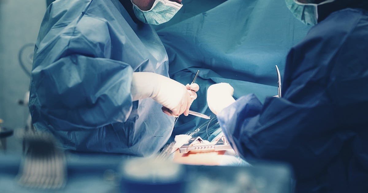
During VATS procedure, cardiothoracic surgeons make 2-3 small incisions (sized 2-3 cm. in diameter) in the side of the chest. Small surgical instruments are inserted to visualize and treat pneumothorax. Possibilities include sewing blisters closed, closing air leaks, or removing the collapsed portion of your lung, which is called a lobectomy. Instead of having a large cut required in the open surgery, VATS does not need breastbone to be divided. As a result, small incisions can significantly lead to less pain, enhanced cosmetic benefit and fewer post-operative complications as well as faster recovery time and shorter hospital.
To minimize chances of recurrence, pleurodesis might be performed. Pleurodesis is defined as a medical procedure in which the pleural space is artificially obliterated. It involves the adhesion of the two pleurae–membranes lining the and enveloping the lungs. This procedure is commonly recommended for individuals who have had repeated episodes of pneumothorax. During the procedure, surgeon irritates the pleural space so that air and fluid can no longer accumulate. Pleurodesis is performed to make the lungs’ membranes stick to the chest cavity. Once the pleura adheres to the chest wall, the pleural space no longer expands and this prevents formation of a future pneumothorax. There are different types of pleurodesis. Mechanical pleurodesis is performed manually by brushing the pleura to cause inflammation. Chemical pleurodesis is another form to deliver chemical irritants such as medical talc to the pleura through a chest tube. The irritation and inflammation cause the lung pleura and chest wall lining to stick together.
Different pleurodesis methods manifest different rates of pneumothorax recurrence. Chances of recurrence is up to 10-20% after chemical pleurodesis while only 1% of pneumothorax recurrence has been observed after mechanical pleurodesis. Therefore, mechanical pleurodesis is superior to other approaches since the recurrence rates are considerably low. Time consumption for operation is approximately an hour, allowing patients to have a shorter stay in the hospital, 2 days on average. Patients can return to their daily life and activities even quicker.
Taking good care after surgery
After surgery, it is highly recommended to:
- Avoid heavy weight lifting. Lifting might cause healing problems to the surgical wound. Complete wound healing process takes approximately 1 week.
- For women with catamenial pneumothorax, hormonal therapy similar to post-menopause treatment is required continuously 6-9 months.
- Lifestyle modification to strengthen lung function include smoke cessation and avoidance of exposure to air pollution such as PM 2.5. Open air activities should be avoided or limited. If necessary, duration of activities must be as short as possible and N95 masks must be worn at all times.

