
Examining heart tissue with 3 Tesla Magnetic Resonance Imaging (MRI)
Translated by AI
Currently, one of the most alarming and increasingly prevalent health hazards is cardiovascular disease. However, with the advancements in medical technology for detecting cardiac abnormalities and reducing impacts on adjacent organs, particularly through the use of Magnetic Resonance Imaging (MRI) technology with a 3 Tesla (3.0 Tesla) magnetic field strength combined with modern software, high-resolution MRI images have been made possible. These images can accurately diagnose abnormalities in heart tissue, heart muscles, and blood vessels.
Heart and Vascular Examination with 3 Tesla MRI
Detecting abnormalities and diagnosing diseases with 3 Tesla (3.0 Tesla) MRI is a technology developed for high-quality and high-resolution diagnosis of heart and vascular abnormalities. This includes identifying diseases of heart tissue abnormalities such as ischemic heart muscle diseases, inflammatory heart diseases, Cardiac Amyloidosis, vascular abnormalities, assessing heart function, heart valve diseases, coronary artery diseases and major blood vessels, and congenital heart diseases before and after treatment in both children and adults. It also includes examinations to assess the risk of coronary artery constriction through pharmacologically induced stress tests (MRI Stress Test with Pharmacologic Induction), as well as exams to observe blood clots in patients with dead heart muscle tissue resulting in low contractility, causing blood in the heart chambers to easily coagulate, among others.
Advantages of 3 Tesla MRI Examinations
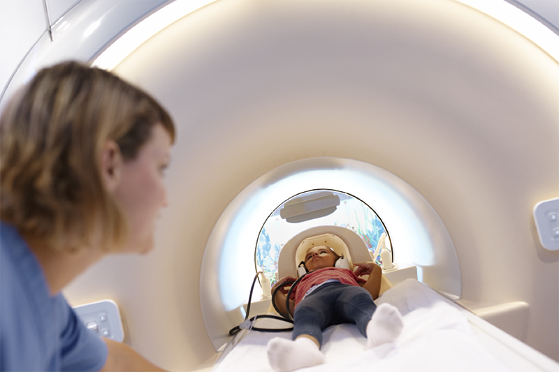
The 3.0 Tesla MRI scan is an alternative for patients who want to avoid the risks of X-ray radiation, ensuring low side effects such as in pregnant patients, patients allergic to contrast media, or those with kidney failure, including elderly patients who cannot hold their breath for long periods, as well as patients without contraindications for MRI examinations. Moreover, the machine has a horizontal cylindrical tunnel that is significantly wider than before, accommodating larger patients and helping reduce claustrophobia. However, for patients with severe claustrophobia, assessments are made on a case-by-case basis, and patients with certain types of artificial heart valves, nerve stimulators, or vascular stents implanted less than 8 weeks prior cannot undergo MRI examinations and are considered for alternate diagnostic methods.
Limits of 3 Tesla MRI Examinations
Although the 3.0 Tesla MRI is a radiation-free tool, there are still limitations to its use, including pregnant or breastfeeding women (in cases where contrast agents are required), patients with metallic medical devices implanted, such as pacemakers, and patients with a kidney filtration rate of less than 30% should consult a nephrologist to assess risks before the examination.
Preparing for a 3 Tesla MRI
Patient preparation before undergoing an MRI includes:
- Fasting from food and water 4 hours before an MRI Stress Test (heart function test using drug stimulation). Other types of MRI exams apart from Stress Tests do not require fasting beforehand.
- Patients undergoing the exam must remove dentures, metal jewelry, etc., as metal components can interfere with magnetic waves.
- Patients who have had metal implants from previous surgeries must inform the staff before entering the examination room.
- Complete bathroom needs before the exam as it may take a long time.
- During the exam, patients must remain still to ensure clear images.
- Patients can signal for assistance if they experience any discomfort, such as chest tightness or difficulty breathing.
MRI technology plays a crucial role in enabling physicians to precisely diagnose diseases and lead to effective and timely treatments by detecting abnormalities in the heart, blood vessels, and heart tissues using magnetic waves (3.0 Tesla MRI)