Magnetic Resonance Imaging of the Heart (Cardiovascular MRI: CMR)
Translated by AI
Cardiac Examination with Magnetic Resonance Imaging
Cardiac examination with Magnetic Resonance Imaging (MRI) is a heart examination that uses magnetic waves instead of X-ray radiation. MRI (Magnetic Resonance Imaging) is a non-invasive tool, which means it does not involve inserting any device into the body. MRI can produce detailed images of the internal structures of different organs and can differentiate between types of tissues. A cardiac MRI can provide both still and moving images of the heart and blood vessels, allowing doctors to use MRI images to diagnose abnormalities in the heart’s structure and function and to plan treatment.
Procedure of MRI
The MRI process usually takes about 30 minutes to 2 hours, depending on the nature of the symptoms and the complexity of the disease. Bangkok Heart Hospital has experienced doctors, nurses, and radiology technicians to care for patients throughout the cardiac MRI examination.
Before the Examination
- Remove jewelry
- Change into hospital-provided garments before the examination
- In some cases, such as patients who need a Stress MRI, which involves administering medication to stimulate the heart through the bloodstream, fasting is required 4 hours before the examination
During the Examination
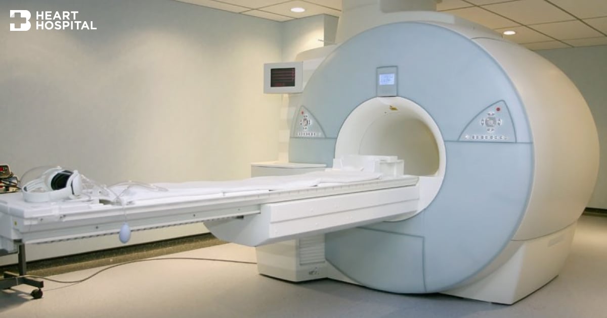
- The MRI machine is tunnel-shaped; patients need to lie flat during the examination
- During the examination, there will be loud noises from the magnetic wave reactions; patients will be given Ear Plugs to block out the noise
- In some cases, an injection of contrast material may be required to enhance the visibility of internal organs
After the Examination
- Resume normal daily activities
The Benefits of Cardiac Examination with Magnetic Resonance Imaging
Cardiac MRI is used for the evaluation and diagnosis of the following diseases:
- Evaluating the anatomical structure, function, and pathology of each part of the heart, such as the heart muscles, heart valves, pericardium, and abnormalities of the blood vessels
- Diagnosing infectious and inflammatory diseases of the heart and blood vessels (Cardiovascular), such as inflammation and infections of the heart muscle or pericardium
- Assessing damage from Coronary Artery Disease, such as restricted blood circulation to nourish the heart muscle and the formation of heart muscle scars or necrosis after a Heart Attack
- Planning treatment for patients with heart and blood vessel diseases
- Monitoring disease progression
- Evaluating changes post-surgery, especially in patients with congenital heart defects
- Evaluating the anatomical structure inside the heart and blood circulation in patients with congenital heart defects
Additionally, cardiac MRI can also be used in conjunction with the results of X-ray or Computed Tomography (CT) for diagnosis, or in some cases, to avoid invasive examinations. In some patients who are unable to undergo CT due to severe seafood allergies, the use of MRI can be particularly beneficial. However, in some cases, the injection of contrast material may be necessary to obtain clearer images of the heart and blood vessels.
Limitations of Diagnostic Examination with Magnetic Resonance Imaging
- Pregnant women
- Individuals with heavy metal prosthetic organs inside their body, such as Cochlear Implants, people who have undergone surgery to place metal clips on bulging blood vessels, individuals with coronary artery stents implanted less than 8 weeks ago, and people with heart pacemakers
- Individuals who cannot lie flat
- Individuals with claustrophobia
- Individuals with metal embedded in their eyes
- Individuals with lead embedded in their body
Bangkok Heart Hospital has extensive experience in diagnosing heart diseases with Magnetic Resonance Imaging (MRI), boasting a multidisciplinary team and specialists capable of diagnosing complicated heart diseases accurately.